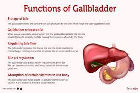
The gallbladder is a small, pear-shaped
organ located just below the liver on the upper right side of the abdomen. Its
main function is to store bile; a digestive fluid produced by the liver, and
releases it into the small intestine when needed to aid in the digestion of
fats.
fa
1. Bile Production:
The liver produces bile continuously, which helps break down fats into smaller
particles that can be easily absorbed. The gallbladder stores this bile until
it is needed for digestion.
2. Bile Composition: Bile
consists of water, bile salts, cholesterol, bilirubin (a waste product), and
other substances. Bile salts play a crucial role in the digestion and
absorption of dietary fats.
3. Gallbladder Issues:
Several conditions can affect the gallbladder. The most common is gallstones,
which are hardened deposits that can form in the gallbladder. Gallstones can
cause pain and other complications, often requiring medical attention.
4. Gallbladder Removal:
In some cases, the gallbladder may need to be removed due to gallstones or
other gallbladder-related problems. This procedure is called a cholecystectomy.
After gallbladder removal, bile flows directly from the liver into the small
intestine, rather than being stored in the gallbladder.
5. Post-Gallbladder Removal:
Most people can live a healthy life without a gallbladder. However, some
individuals may experience changes in their digestion, such as difficulty
digesting fatty foods. Eating smaller, more frequent meals and avoiding
high-fat meals can help manage these symptoms.
Diagnostic
Tools
There are several diagnostic tools and
tests that healthcare professionals use to diagnose cholelithiasis
(gallstones). These diagnostic methods include:
1. Medical History and Physical Examination:
The doctor will ask about your symptoms, medical history, and any risk factors
associated with gallstones. They will also perform a physical examination to
assess for signs of gallbladder disease.
2. Imaging Tests:
· Ultrasound:
Ultrasound imaging is the most commonly used and non-invasive method to
diagnose gallstones. It uses sound waves to create images of the gallbladder
and can detect the presence, size, and location of gallstones.
· Computed
Tomography (CT) Scan: A CT scan can provide detailed
cross-sectional images of the abdomen, including the gallbladder and
surrounding structures. It may be used if ultrasound results are inconclusive
or to evaluate complications related to gallstones.
· Magnetic
Resonance Cholangiopancreatography (MRCP): MRCP
combines magnetic resonance imaging (MRI) with specialized techniques to create
images of the bile ducts and gallbladder. It can help visualize gallstones and
identify any associated complications.
3. Blood Tests:
Blood tests may be performed to assess liver function, check for signs of
inflammation or infection, and evaluate the levels of specific enzymes and
bilirubin in the blood. These tests can help determine the extent of
gallbladder or bile duct involvement.
4. Endoscopic Retrograde
Cholangiopancreatography (ERCP): ERCP is
primarily a therapeutic procedure but can also aid in diagnosing gallstones.
During ERCP, a flexible tube with a camera is passed through the mouth,
esophagus, and stomach to the small intestine. Contrast dye is then injected
into the bile ducts, and X-ray images are taken to identify gallstones or other
abnormalities.
F
Risk Factors
1. Age and Gender:
Cholelithiasis is more common in individuals over the age of 40. Women are also
at a higher risk, especially those who have had multiple pregnancies, hormone
replacement therapy, or are taking oral contraceptives.
2. Obesity:
Obesity is a significant risk factor for developing gallstones. It can lead to
increased cholesterol levels in bile and decreased gallbladder motility,
contributing to stone formation.
3. Rapid Weight Loss:
People who lose weight quickly, such as through crash diets or bariatric
surgery, have an increased risk of developing gallstones. Rapid weight loss can
disrupt the balance of bile salts, cholesterol, and other substances in the
bile, promoting stone formation.
4. Diet:
A high-fat, low-fiber diet may increase the risk of cholelithiasis. Consuming
excessive amounts of cholesterol-rich foods and refined carbohydrates can
contribute to the formation of gallstones.
5. Sedentary Lifestyle:
Lack of physical activity and prolonged periods of immobility can increase the
risk of gallstone formation.
6. Family History:
A family history of gallstones increases the likelihood of developing
cholelithiasis. There may be genetic factors that predispose individuals to
gallstone formation.
7. Certain Medical Conditions:
Certain medical conditions, such as cirrhosis of the liver, Crohn's disease,
and metabolic disorders like diabetes, are associated with an increased risk of
gallstone formation.
8. Medications:
Certain medications, such as hormone replacement therapy, oral contraceptives,
and cholesterol-lowering drugs, may increase the risk of gallstones.
Harms
Cholelithiasis, or the presence of gallstones in the gallbladder, can lead to various harms and complications. Some of the potential harms associated with cholelithiasis include:
1. Gallstone Obstruction:
Gallstones can block the bile ducts, preventing the flow of bile from the
gallbladder to the small intestine. This can cause intense pain in the upper
abdomen, known as biliary colic. The pain may be accompanied by nausea,
vomiting, and jaundice.
2. Cholecystitis:
If a gallstone blocks the cystic duct (the duct that connects the gallbladder
to the common bile duct), it can lead to inflammation of the gallbladder, known
as cholecystitis. Cholecystitis can cause severe abdominal pain, fever, and
infection.
3. Choledocholithiasis:
Gallstones can also pass from the gallbladder into the common bile duct. This
condition, called Choledocholithiasis, can obstruct the bile flow and lead to
jaundice, abdominal pain, and potentially severe complications such as
pancreatitis.
4. Pancreatitis:
In some cases, gallstones can travel from the common bile duct and obstruct the
pancreatic duct, causing inflammation of the pancreas. This condition, known as
gallstone pancreatitis, is a serious and potentially
life-threatening complication.
5. Biliary Infections:
When the bile flow is obstructed by gallstones, it can create an environment
that is favorable for the growth of bacteria, leading to biliary infections
such as cholangitis. These infections can cause symptoms like
fever, abdominal pain, and jaundice, and require immediate medical attention.
6. Biliary Fistula or Perforation:
In rare cases, gallstones can erode through the gallbladder or bile ducts,
creating abnormal connections (fistulas) between the gallbladder, bile ducts,
or other nearby organs. This can result in infection, abscess formation, or
even perforation of the gallbladder.
7. Increased Risk of Gallbladder Cancer:
Although the majority of gallstones do not lead to cancer, long-standing
gallstones may increase the risk of developing gallbladder cancer, a relatively
rare but serious malignancy.
Treatment Options:
The treatment options for cholelithiasis
(gallstones) depend on the presence of symptoms, the size and number of
gallstones, and the overall health of the individual. The treatment approaches
for cholelithiasis include:
1. Watchful Waiting:
If gallstones are small and asymptomatic, a "wait-and-see" approach
may be recommended. This involves monitoring for symptoms and complications
while making lifestyle modifications to prevent symptom development.
2. Medications:
Medications are generally not effective in dissolving gallstones, but certain
medications can be prescribed to manage symptoms or dissolve specific types of
gallstones, such as:
· Ursodeoxycholic
acid (UDCA): UDCA can be used to dissolve
cholesterol gallstones in some cases, particularly for those who cannot undergo
surgery.
· Oral
dissolution therapy: This involves taking medications
that help break down cholesterol gallstones over a period of months or even
years. This approach is typically used for patients with small cholesterol
stones and intact gallbladders.
3. Surgery:
· Cholecystectomy:
The most common and definitive treatment
for symptomatic gallstones is the surgical removal of the gallbladder, known as
cholecystectomy. This can be done through two main approaches:
· Laparoscopic
Cholecystectomy: It is a minimally invasive
procedure performed using small incisions and a laparoscope, which allows the
surgeon to visualize and remove the gallbladder.
· Open
Cholecystectomy: This traditional surgical approach
involves a larger incision in the abdomen to remove the gallbladder. It is
typically reserved for cases with severe inflammation, scarring, or
complications.
· Endoscopic
Retrograde Cholangiopancreatography (ERCP):
ERCP is a procedure used to remove gallstones that have migrated from the
gallbladder into the common bile duct. A specialized endoscope is inserted
through the mouth, down the esophagus, and into the bile ducts to remove or break
down the stones.
Percutaneous Cholecystectomy: This
procedure is typically performed in individuals who are not candidates for
surgery due to high surgical risks. It involves inserting a tube through the
skin and into the gallbladder to drain bile and relieve symptoms temporarily.
Fahad
The choice of
treatment depends on various factors, such as the presence of symptoms, the
size and type of gallstones, and the individual's overall health. It is
important to consult with a healthcare professional to determine the most
suitable treatment option for cholelithiasis based on the individual's specific
circumstances.


